Migration activity analysis
|
|
The animal-cell capability of active migration has been the object of interest of many research laboratories all over the world, as the process plays a substantial role in many physiological processes and is related to many pathological conditions. The active migration of cells takes place during the embryonic development, wound healing, defensive reactions of immunological system, cancer cells metastasis, and other processes. The Department of Cell Biology is fully equipped with the apparatus and software to register and quantitatively analyse cell movement. The microscopic image analysis system consists of a microscope, a heating chamber which keeps the temperature at 37 °C, a video camera, and a computer with image acquisition software and time-lapse software. The application of computer systems in cell migration research involves determining the changes in cell position during the experiment. The most frequent method used to determine the cell position consists in marking its centroid position.
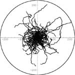
Plot-creation procedures from ‘‘Mathematica'' have been used for the visualisation of results. The programme permitted the transfer of all cell trajectories from each experiment to the circular diagrams as suggested by Erickson and Nuccitelli [1984], Gruler and Nuccitelli [1991], Nuccitelli et al. [1993], Nishimura et al. [1996], and Sheridan et al. [1996]. We have used the method proposed by Erickson and Nuccitelli [1984] and Gruler and Nuccitelli [1991] to illustrate the orientation of cell movement, but instead of final cell displacements we have used full cell trajectories with the starting positions of cells at the origin of the coordinate axes. The x axis is the axis of electric field direction. Pooled data from experiments have been used for the statistical analysis of the parameters measured. Histograms of intersegmental and directional angles distribution have been prepared as described by Gail [1973], Gruler [1984], Gruler and Nuccitelli [1991].
The measured parameters include:
- the total length of cell displacement from the starting point to the final cell position,
- the total length of cell trajectory, i.e., the ‘‘true'' length of cell trajectory; the trajectory of a cell has been considered as a sequence of ‘‘n''straight-line segments, each corresponding to the cell centroid translocation within one time interval between two successive images,
- the ratio of cell displacement to cell trajectory length defined as the coefficient of movement efficiency (CME),
- average speed of cell locomotion (calculated from the length of cell path in a given time),
- average speed of cell displacement,
- angles between successive segmental displacements.
- average directional cosines, indicating directionality of cell movement,
- average instantaneous velocity.
|
|
Besides determining the cell-movement trajectory, the cell-movement analysis uses methods based on the analysis of changes in the shapes of the migrating cells, that is the analysis of their contours and determination of the parameter which describes the ratio of new surface covered by pseudopodium moving in the movement direction to the overall surface of the cell.